
It seems like you might be asking about “angiography” (possibly due to a typo or miscommunication). Angiography is a medical imaging technique used to visualize the inside of blood vessels and organs. It is commonly used to identify blockages, aneurysms, and other issues in blood vessels, particularly in the context of heart disease, brain conditions, and other vascular disorders.
What is Angiography?
Angiography is a diagnostic procedure that involves injecting a contrast dye into the blood vessels to make them visible on X-rays or other imaging techniques. The contrast dye helps to outline blood vessels and highlight any abnormalities in the flow or structure of the vessels.
Types of Angiography
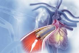
- Coronary Angiography (Heart Angiography):
- Used to examine the coronary arteries of the heart.
- This type of angiography helps detect blockages or narrowing of the coronary arteries, which can lead to heart attacks.
- It is typically done if a patient has symptoms like chest pain or shortness of breath, which may indicate coronary artery disease (CAD).
- Cerebral Angiography (Brain Angiography):
- Used to visualize the blood vessels in the brain.
- It can identify issues like aneurysms, arteriovenous malformations (AVMs), and stenosis (narrowing of the blood vessels).
- It is typically done for patients with neurological symptoms like sudden severe headaches, dizziness, or stroke-like symptoms.
- Peripheral Angiography:
- Used to examine the blood vessels in the legs and arms.
- It can help detect peripheral artery disease (PAD), which causes narrowing of the arteries in the limbs and can lead to pain, ulcers, and difficulty walking.
- Pulmonary Angiography:
- Used to evaluate the blood vessels in the lungs.
- It is mainly used to detect pulmonary embolism (PE), a blockage in the pulmonary arteries caused by blood clots.
- Renal Angiography:
- Used to look at the blood vessels of the kidneys.
- This test is often used to identify renal artery stenosis (narrowing of the arteries supplying blood to the kidneys), which can cause high blood pressure or kidney failure.
Procedure:
- Preparation:
- The patient may be asked to fast for a few hours before the procedure.
- The doctor may administer a mild sedative to help the patient relax.
- Contrast Injection:
- A catheter (thin flexible tube) is inserted into a blood vessel, typically in the groin or arm.
- The catheter is carefully guided to the area of interest, and a contrast dye is injected through the catheter into the blood vessels.
- The dye helps to make the blood vessels visible on X-ray, CT scan, or MRI images.
- Imaging:
- Once the dye is injected, a series of X-rays or other imaging scans are taken to capture the flow of the dye through the blood vessels.
- Post-procedure care:
- The catheter is removed, and pressure is applied to the insertion site to stop any bleeding.
- The patient may need to lie still for a while to prevent complications.
- The patient is monitored for any immediate reactions to the contrast dye or sedation.
Why is Angiography Done?
Angiography is typically performed to:
- Identify Blockages: To detect any narrowing or blockages in blood vessels that could lead to conditions like heart attacks or strokes.
- Guide Treatment: It helps doctors plan for procedures like angioplasty (to open blocked arteries) or stent placement (to keep an artery open).
- Diagnose Vascular Conditions: To diagnose conditions like aneurysms, arteriovenous malformations, or vessel malformations that could cause serious health problems.
- Evaluate Blood Flow: It helps assess the blood flow to organs like the heart, brain, and kidneys.
Risks of Angiography
While angiography is generally a safe procedure, there are some risks associated with it, including:
- Allergic Reactions: Some people may be allergic to the contrast dye.
- Bleeding: The catheter insertion site (usually the groin or arm) can sometimes bleed.
- Infection: There is a small risk of infection at the catheter insertion site.
- Kidney Damage: The contrast dye can sometimes affect kidney function, particularly in people with pre-existing kidney conditions.
- Radiation Exposure: X-ray angiography involves exposure to radiation, though the amount is typically very low and well-controlled.
Advantages of Angiography:
- Accurate Diagnosis: It provides detailed images of blood vessels, helping doctors accurately diagnose vascular issues.
- Guides Treatment: It helps in planning treatments like angioplasty, stent placement, or surgery.
- Non-invasive Option: In many cases, angiography can be performed with minimal invasion, especially if a catheter is used through a small incision.